

Shah's Gastro, Cancer & Robotic Surgery Centre · Ahmedabad & Gandhinagar
One measurement taken at endoscopy settles which operation is done. Apollo Hospital, Bhat · Gota OPD, Ahmedabad.
A tumour sitting where the food pipe joins the stomach is not one disease with one operation. It is classified by where its centre sits in relation to the anatomical cardia — the exact point at which the food pipe ends and the stomach begins — and that classification, called the Siewert type, decides whether the surgeon removes most of the food pipe and pulls the stomach up into the chest, or removes the whole stomach and joins the food pipe to a loop of small bowel. Dr Harsh Shah’s position is that this measurement belongs in the endoscopy report in centimetres, is confirmed on the scan, and is taken at a tumour board before any family is told what operation is planned.
Call +91-63555-64601 · Send your reports to Dr Harsh Shah’s office on WhatsApp
Shah’s Gastro, Cancer & Robotic Surgery Centre. Cancers at the junction of the food pipe and the stomach have become commoner over the last two decades, and they are the one site in the digestive tract where two different specialties can each look at the same tumour and describe a different operation. The disagreement is almost never about skill. It is about where the tumour’s centre was measured from.
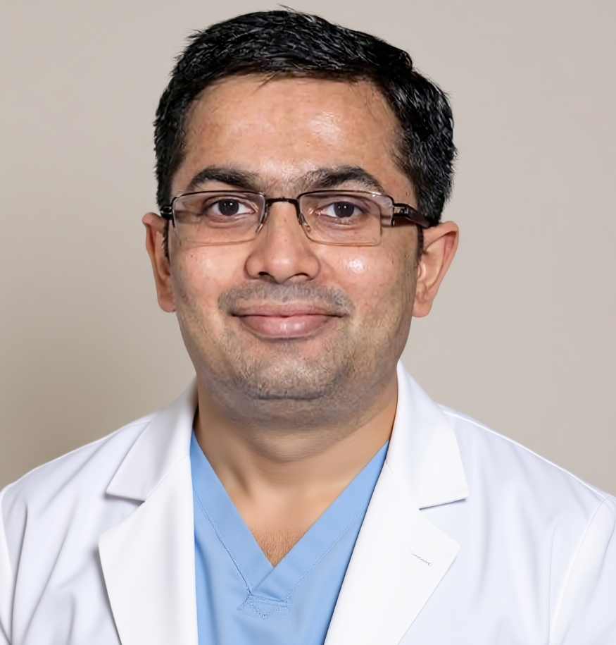
Dr Harsh Shah
MS, MCh (Surgical Gastroenterology), DrNB (Surgical Gastroenterology)
Registered with Gujarat Medical Council, Reg. No. G-38738
Food Pipe Cancer Surgeon · Apollo Hospital, Bhat, Gandhinagar · Gota OPD, Ahmedabad
Google rating 4.89 from 216 reviews at the Gota clinic, and 4.94 from 17 reviews at Apollo, Bhat.
The food pipe and the stomach drain to different sets of lymph nodes. The lower food pipe drains upwards into the chest; the upper stomach drains downwards into the abdomen. A tumour straddling the join can drain in both directions, and the operation has to go where the drainage goes. That is why the position of the tumour’s centre, measured in centimetres from the anatomical cardia, changes the whole operation rather than a detail of it.
Dr Harsh Shah repeats the endoscopy himself in a good number of these patients, not out of distrust of the first endoscopist, but because the surgeon who will have to define the upper margin in theatre benefits from having seen the tumour’s upper edge with his own eyes. The same applies to the lower edge, which decides how much stomach can be kept.
Siewert type I sits mainly above the join and is treated as a food pipe cancer — the food pipe is removed and the stomach is reshaped into a tube and brought up to replace it. Type III sits mainly below the join and is treated as a stomach cancer — the whole stomach is removed together with the lowest part of the food pipe. Type II straddles the join, and is the type on which units genuinely differ; Dr Harsh Shah’s practice follows the guidance that favours removing the food pipe rather than extending a stomach operation upwards.
| Where the centre of the tumour sits | Siewert type | The operation this points to |
|---|---|---|
| Above the anatomical cardia, in the lower food pipe | Type I | Oesophagectomy — the food pipe is removed with its chest lymph nodes, and the stomach is made into a tube and joined to what remains of the food pipe |
| Straddling the join, just above to just below the cardia | Type II | Oesophagectomy preferred over an extended stomach operation, so that the nodes in the chest are cleared as well as those in the abdomen |
| Below the cardia, in the upper stomach | Type III | Extended total gastrectomy — the whole stomach is removed along with the lowest segment of the food pipe, and the food pipe is joined to a loop of small bowel |
| Tumour reaching the windpipe, the main airways or the aorta | Any type | Surgery is not the first step, and may not be part of the plan at all; chemotherapy with radiotherapy takes its place |
| Disease already in the liver, lungs or the lining of the abdomen | Any type | The operation does not help; drug treatment and relief of swallowing become the plan |
One line on that table causes avoidable distress. A type II tumour is not a borderline case in the sense of being uncertain — it is a defined type with a defined recommended approach, and being told “it is in between” is a description of the anatomy, not of the plan.
Before any operation is planned, the endoscopy report should state the distance from the front teeth to the upper edge of the tumour, to the lower edge, and to the anatomical cardia; the length of tumour; how much of the circumference it occupies; and whether the scope passed through. Without those numbers the Siewert type cannot be assigned, and without the type nobody can say which organ is to be removed.
Alongside that, a contrast scan of the chest, abdomen and pelvis is done, and a PET-CT is added for every patient in whom an operation is being considered, because it finds disease elsewhere in a meaningful number of people and spares them an operation that could not have helped. Endoscopic ultrasound is used where it will change the decision, chiefly in sampling a doubtful lymph node.
Two tests are often skipped and should not be. Lung function testing with gas transfer, and an echocardiogram, are done before any operation involving the chest; they, and not the patient’s age, decide fitness for the thoracic part. And where the tumour is higher up, the airway is inspected with a bronchoscope, because a tumour pressing on the main airways changes the plan entirely.
The biopsy result splits these patients into two pathways that look nothing alike. Adenocarcinoma at the junction is usually treated with chemotherapy before and after surgery. Squamous cell carcinoma of the food pipe is usually treated with chemotherapy and radiotherapy together before surgery. Giving one pathway’s treatment to the other patient is the commonest avoidable error at this site.
Most junctional tumours turn out to be adenocarcinoma, and for a fit patient with locally advanced disease the current standard is a course of combination chemotherapy before the operation and the same chemotherapy again after it, with surgery in the gap. That schedule came out ahead when it was compared directly against chemotherapy with radiotherapy in a randomised trial of junctional and food pipe adenocarcinoma, which is why Dr Harsh Shah offers it as the default for this histology.
Squamous cell carcinoma behaves differently and responds strikingly well to radiotherapy given with chemotherapy. For those patients the sequence is chemotherapy with radiotherapy first, then surgery roughly six to eight weeks after it finishes, once the tissues have settled enough to be operated on safely.
What happens afterwards is also decided by the pathologist, on the specimen this time. Where the disease has disappeared completely, no further treatment is added and surveillance begins. Where disease remains after chemotherapy with radiotherapy and a complete removal, a year of immunotherapy is offered, started within a defined window rather than whenever an appointment happens to be free. Where the patient was on the chemotherapy pathway, the remaining cycles are completed.
For an adenocarcinoma straddling the junction, Dr Harsh Shah usually begins with a staging laparoscopy: a short procedure through two or three small cuts, with a camera, to inspect the lining of the abdomen before committing to the full operation. Deposits too small to appear on any scan are found often enough at this site to make the few minutes worth taking.
Washings are taken from the abdomen at the same time and examined for cancer cells. A positive washing in the absence of any visible deposit is a genuine finding that changes the sequence of treatment, and it is one of the reasons the look is done at all.
An oesophagectomy removes the affected length of the food pipe together with the lymph nodes in the chest and the upper abdomen that drain it. The stomach is then made into a narrow tube, brought up through the chest, and joined to the remaining food pipe — inside the chest for a lower tumour, or in the neck for a higher one.
A proper node clearance is not an optional extra. The pathologist needs a real number of nodes to be able to say whether the disease had spread, and a handful of nodes cannot answer that question. Where the tumour is an adenocarcinoma, a clear length of normal food pipe above the tumour is also taken, and the margin is checked on frozen section during the operation when there is any doubt.
Dr Harsh Shah performs this operation by a minimally invasive route — laparoscopic or robotic — wherever the anatomy allows, because the chest complications that dominate recovery after this operation are fewer when the chest is not opened widely. Where the tumour is bulky, or the tissues are heavily scarred after radiotherapy, he says at consent that an open operation may be the safer course rather than promising a keyhole procedure the findings may not allow.
For a tumour centred below the join, the whole stomach is removed together with the lowest segment of the food pipe and the lymph nodes of the upper abdomen. The food pipe is then joined directly to a loop of small bowel, which becomes the route food takes. The spleen is not removed routinely, and is taken only where the tumour or its nodes make it necessary.
The reason the operation reaches upwards into the lower chest is margin. A tumour that has come up to the join has usually crept further up the food pipe wall than it appears, so a length of normal food pipe above it is taken and, where there is doubt, checked under the microscope while the patient is still asleep. If that check comes back involved, more food pipe is removed at the same sitting rather than at a second operation later.
Eating after a total gastrectomy changes permanently and this is counselled before, not after. The reservoir is gone, so meals become small and frequent for life, sweet liquid taken quickly on an empty stomach can cause faintness and cramps, and vitamin B12 has to be replaced by injection because the stomach lining that absorbed it is no longer there.
★★★★★
“We are very thankful to Dr. Harsh Shah for treating and operating on my father for gastric (stomach) cancer. From the first consultation to the surgery and follow-up, he guided us with great care and explained everything clearly. My father is doing well now, and we are extremely grateful for the treatment and support provided by Dr. Harsh Shah and his team. He is a highly experienced and compassionate doctor. We would definitely recommend Dr. Harsh Shah to anyone looking for an experienced doctor for stomach cancer”
★★★★★
“My father has carcinoma of stomach ,Harsh sir did radical gastrectomy and he is recovering very nicely under Dr.Harsh's observation ,he listen each and every complain calmly and with patience and make patient very comfortable during treatment , during procedure and during stay at hospital. we are satisfied with the treatment and very confident about the fully recovery of disease as we are under treatment of Dr.Harsh & team.Thank you Dr.Harsh & his whole team and staff.”
★★★★★
“Dr Harsh shah's dedication and commitment to his patients health are commendable. He always provides comprehensive explanations and ensures that we understood our treatment plans.Dr harsh shah is best stomach cancer surgeon in Ahmedabad Gujarat. Thank u so much sir for all support and help.”
Moh. Ahsan · Google review
tina parmar · Google review
Haresh Chavda · Google review
What is my Siewert type, and what measurement was it based on? The answer should be a number in centimetres from a named report, not an impression.
Which organ are you removing — the food pipe or the stomach — and what takes its place? Ask for the answer in those words. It is the question most often left unanswered.
What does my biopsy say the tumour is made of, and which pathway does that put me on? Adenocarcinoma and squamous cell carcinoma are treated differently before surgery, and the answer should name which one you have.
Has my case been discussed at a tumour board, and what did that meeting decide? Every junctional tumour should go to that meeting before treatment starts.
What would make you stop the operation? Finding disease in the lining of the abdomen, or an upper margin that cannot be cleared, are the honest answers.
A second opinion earns its keep in three situations here: when you have been told the whole stomach must come out but nobody has stated your Siewert type; when surgery is being planned before any chemotherapy in a locally advanced tumour; and when you have been refused an operation on the basis of a scan alone, without a staging laparoscopy having been considered.
One. Book your consultation. Bring the endoscopy report with the measurements, the biopsy slides and blocks, all scans on a disc rather than as printed films, and a list of your current medicines.
Two. Staging and assessment — a contrast scan and PET-CT, an endoscopy repeated by the surgeon where the upper edge matters, lung function with gas transfer, an echocardiogram, and a nutrition review with a dietitian.
Three. Your case goes to the tumour board, and a written plan comes back naming the Siewert type, the histology, whether treatment starts with chemotherapy or with chemotherapy and radiotherapy, and which operation follows.
Four. Surgery at Apollo Hospital, Bhat — a staging laparoscopy first where it is indicated, then the planned operation, with the recovery pathway, the feeding plan and the chest physiotherapy explained to your family beforehand.
Five. Histology review at about two weeks, the decision on treatment after surgery, a dietitian plan in writing, and a surveillance schedule you take home on paper.
There is no single figure for an operation at the junction, and any page offering one is guessing. What moves it is which operation is done, whether the chest is opened or the procedure is done by a minimally invasive route, how long the stay runs, the room category, whether intensive care is needed, and whether chemotherapy or radiotherapy is given before and after.
The office prepares a written estimate before admission, so that you are comparing a document rather than a conversation. Cashless admission is arranged where your policy allows it, and the pre-authorisation paperwork is prepared for you. Ask what the estimate excludes, and who your point of contact is if the hospital and the insurer disagree.
Two practical points specific to this situation. Treatment before surgery means two separate claims months apart, so keep the biopsy report and the tumour board note together — they are the documents that link the two. And if a feeding tube is planned at the time of surgery, ask for it to appear in the estimate.
Both may be describing the correct operation for a different Siewert type. Ask each of them what type they assigned and what measurement they used. Once the type is agreed, the disagreement usually disappears.
The stomach is made into a narrow tube and brought up to take the place of the food pipe, and it is joined to what remains of it. Swallowing works again, in smaller mouthfuls and with the head raised at night. The join can narrow over time and is opened with a short endoscopic procedure when it does.
Yes, in suitable patients. Robotic and laparoscopic routes are used at this site because the chest complications that dominate recovery are fewer when the chest is not opened widely. Bulky tumours and tissues scarred by radiotherapy may still be safer open.
Treatment given before surgery shrinks the tumour away from the structures around it, treats disease too small to see, and is tolerated far better before an operation than after one. For junctional adenocarcinoma this sequence gave longer survival than the alternative when the two were compared directly in a randomised trial.
Not reliably. Difficulty in swallowing appears only once a good part of the food pipe's width is blocked, so a tumour can be well established while swallowing is still comfortable. The stage is decided by the scans and the endoscopy, not by symptoms.
Shah’s Gastro, Cancer & Robotic Surgery Centre — Gota, Ahmedabad
Consultation, review of the endoscopy and biopsy reports, second opinions, surveillance.
Google: Dr Harsh Shah · 4.89 from 216 reviews
Directions to the Gota clinic
Apollo Hospital — Bhat, Gandhinagar
Admission, surgery and inpatient care.
Google: Dr Harsh Shah – Robotic GI Surgeon · 4.94 from 17 reviews
Directions to Apollo, Bhat
Patients are referred for junctional and food pipe cancer from across Gujarat, southern Rajasthan and western Madhya Pradesh. Send the endoscopy report with its measurements and the biopsy report ahead of the appointment, so that the Siewert type can be assigned before you travel.
More on Dr Harsh Shah’s work at this site: food pipe cancer treatment · stomach cancer surgery · robotic cancer surgery.
Swasth Parivar: every week, practical advice on recovery, diet and warning signs. No advertising. Stop any time.
Your number is used for these updates and for the clinic to reach you. It is never sold or passed on. To stop, reply STOP on WhatsApp.